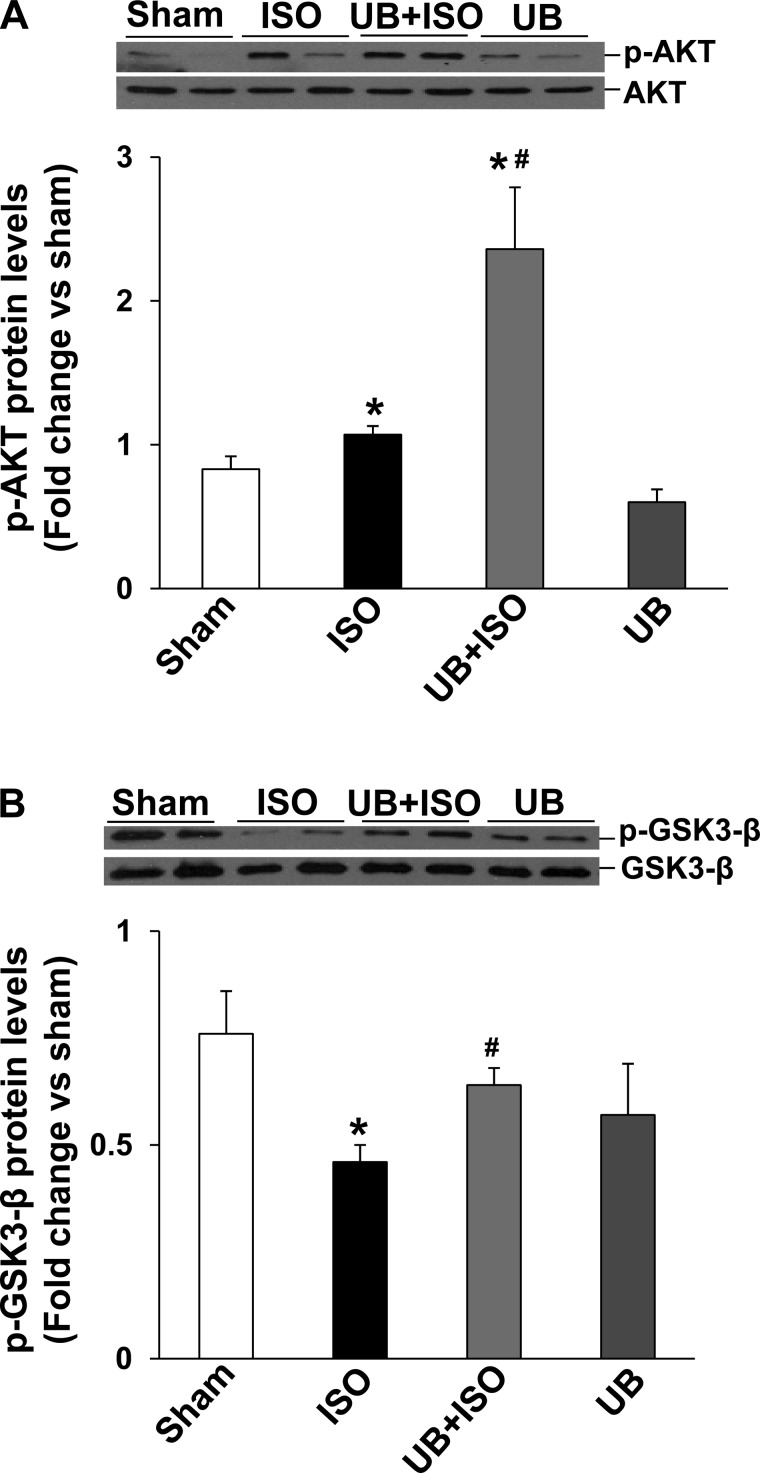
Fig. 3.
Activation of Akt and GSK-3β in the heart. Total left ventricular (LV) lysates (70 μg) were analyzed by Western blot using phosphospecific (p)-Akt (Ser473), total Akt, phosphospecific GSK-3β (Ser9) and total GSK-3β antibodies. A and B, bottom, exhibit mean data normalized to total Akt or GSK-3β. A: phosphorylation (activation) of Akt. *P < 0.05 vs. sham; #P < 0.05 vs. ISO; n = 6. B: phosphorylation (inactivation) of GSK-3β. *P < 0.05 vs. sham; #P < 0.01 vs. ISO; n = 4–7.

