Fig. 7.
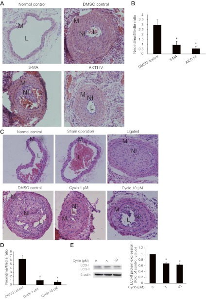
Neointima formation of mouse carotid arteries is inhibited by 3-MA, AKTI IV, and cyclopamine. Neointimal lesions were generated through ligation of mouse common carotid arteries as described in methods. 3-MA, AKTI IV, and cyclopamine were applied through PLF-127 gel. DMSO was the vehicle control for cyclopamine. A and B: representative micrographs of H&E staining (A) showing the effects of 3-MA and AKTI IV on the development of neointima (original magnification, ×200). The sizes of the lesions (B) were calculated as the ratio of the neointima area to that of the media (*P < 0.01 vs. DMSO control; n = 3). C and D: representative micrographs of H&E staining showing the effects of cyclopamine on the development of neointima (C; original magnification, ×200). The sizes of neointimal lesions were calculated as the ratio of the area of the neointima to that of the media (D; *P < 0.01 vs. DMSO control, n = 3). E: Western blot results (left) showing the effects of cyclopamine on LC3-II conversion in neointimal lesions. The intensity of bands was quantified and normalized to the DMSO control (right, *P < 0.01, n = 3).

