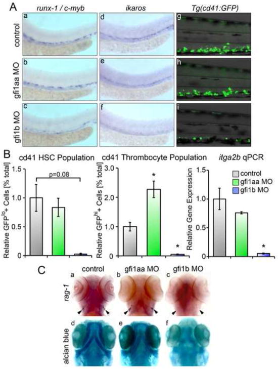
Figure 5. Loss of gfi1b silences definitive HSC.
(A) Loss of gfi1b silences runx-1, c-myb and ikaros expressing HSC in the AGM at 36 hpf relative to matched controls (a–f). Loss of gfi1b also reduces expression of GFP+ cd41 cells in Tg(cd41:eGFP) embryos at 96 hpf (d–f). (B) FACS of Tg(cd41:eGFP) embryos at 96 hpf injected with MO targeting either gfi1aa or gfi1b reveals a marked reduction in the GFPlo and GFPhi populations in gfi1b morphants (mean ± SE, t test, *p < 0.05, n = 3). qRT-PCR of gfi morphants shows significantly reduced expression of itga2b (cd41) in gfi1b morphants at 96 hpf (mean ± SE, t test, *p < 0.05, n = 3). (C) Loss of gfi1b silences rag-1 expressing thymic lymphocytes, while loss of gfi1aa has no effect on lymphopoiesis in the thymic anlage (a–c). (d–f) Embryos were stained with Alcian Blue to delineate the morphologic architecture of the jaw cartilages. gfi1aa morphants have normal jaw cartilage development compared with wild type controls (d,e), while gfi1b morphants show dysplastic development of the jaw cartilage (f).

