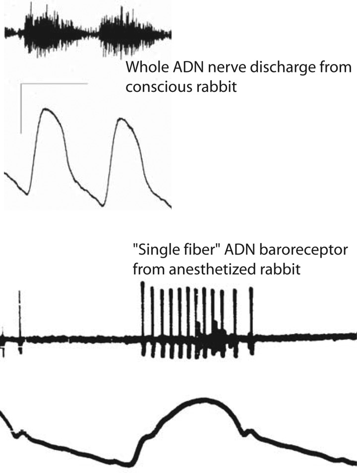
Fig. 1.
Whole nerve activity and single fiber nerve activity from the aortic depressor nerve (ADN). Top: a whole ADN activity recorded for two cardiac cycles from a conscious rabbit while occlusion of the descending aorta forced arterial pressure higher. Bottom: recorded activity from a thin, split fiber divided from the ADN of an anesthetized rabbit. Whole nerve recording registers activity from a nerve trunk containing thousands of axons but reflects electrical signal interactions from an unknown number of these axons (14). The single fiber recording shows the phasic activation of a regularly discharging aortic baroreceptor whose instantaneous frequency encodes and reports details of the arterial pressure typical of myelinated, A-type baroreceptors. A cluster of small amplitude spikes likely from C-type baroreceptors fires sparsely only at the peak of systole.

