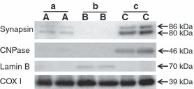
Figure 4.

Representative western blots showing the contamination by other cellular fractions in the isolated mitochondria from PND 9 mouse brain using different protocol A (a), protocol B (b) and protocol C (c). Synapsin, synapse marker; CNPase, myelination marker; Lamin B, nuclear marker; COX I, loading control.
