
Figure 5.
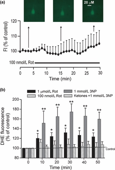
Changes in ROS levels after MRC inhibition. (a) No changes in DCF fluorescence intensity (FI) were seen in CA1 pyramidal neurons exposed to 100 nmol/L Rot over 30 min. (b) In the control group, pyramidal neurons were perfused with aCSF solution containing dithroethidium (DHE), and only a slight increase in DHE intensity was seen over time. Rot evoked a dose-dependent increase in DHE fluorescence intensity, but 100 nmol/L Rot did not evoke a significant change in DHE intensity compared to control. One mmol/L 3-NP prominently enhanced reactive oxygen species (ROS) among the three groups, whereas ketone application largely suppressed ROS generation induced by 3-NP. Values represent mean ± SEM. One way anova followed by Tukey test; *, p < 0.05; **, p < 0.01.