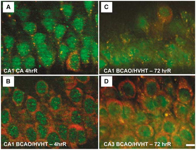
Figure 6.

Stress granules in ischemic and reperfused CA1. (A) At 4 h reperfusion after a 10 mins cardiac arrest (CA), small ribosomal protein S6 (red) is exclusively localized in SGs, as marked by cytoplasmic colocalization with TIA-1 (green). (B) After 4 h reperfusion after 10 mins forebrain ischemia induced by bilateral carotid artery occlusion and hypovolemic hypotension (BCAO/HVHT) to 40mm Hg, CA1 pyramidal neurons resemble controls with robust free S6 staining, and a few cytoplasmic SGs. (C) At 3 days reperfusion after 10 mins BCAO/HVHT, CA1 pyramidal neurons show S6 to be exclusively localized to SGs, similar to the 4 h cardiac arrest animals. (D) However, at 3 days reperfusion after 10 mins BCAO/HVHT, pyramidal neurons of CA3 resemble controls, with robust free S6 staining, and a few cytoplasmic SGs. In (A) and (C), pyramidal layer interneurons in CA1, identified by their large cytoplasm, show staining patterns like controls, indicating that the alteration of SGs is exclusively localized to pyramidal neurons and not interneurons in CA1, consistent with known patterns of protein synthesis inhibition and cell death. Immunohistochemistry was performed as described in Kayali et al (2005). Scale bar in (D) is 10 μm and applies to all four panels.
