Figure 2.

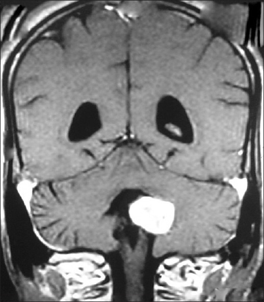
MRI brain with gadolinium, coronal view, showing welldefined tumor in lateral recess with part of the tumor free in 4th ventricle

MRI brain with gadolinium, coronal view, showing welldefined tumor in lateral recess with part of the tumor free in 4th ventricle