Figure 1.

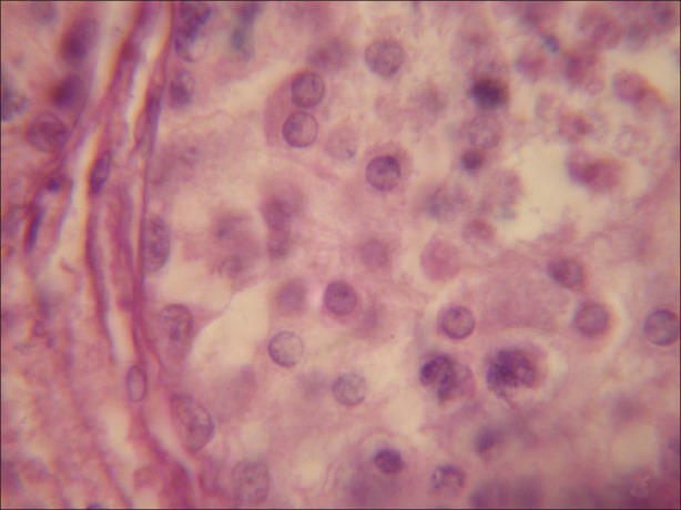
Testicular tissue exhibiting normal structure of seminiferous tubule, having normal Leydig cells, Sertoli cells and different types of germ cells. (×1000)

Testicular tissue exhibiting normal structure of seminiferous tubule, having normal Leydig cells, Sertoli cells and different types of germ cells. (×1000)