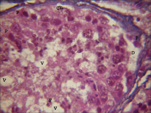
Figure 2.

Light microphotograph of testicular portion treated with atrazine (100 nmolml-1) without antioxidant after 1 hour, showing initiation of degeneration in all types of cells. Pycnosis (P) and chromolysis (Ch) were clearly visible. Vacuoles of varying sizes and shapes were observed. Basal lamina was detached due to the exposure of atrazine (D). (×1000)
