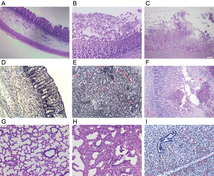
Figure 3.
Histopathology images from antibody-treated piglets inoculated with Clostridium difficile. A, Submucosal edema with intact mucosa, minimal neutrophilic infiltration, and no formation of pseudomembranes in the spiral colon of a piglet treated with polyclonal anti-TcdB. B, Extensive neutrophilic infiltration of the mucosa with pseudomembrane formation in the spiral colon of a piglet treated with polyclonal anti-TcdA. C, Neutrophilic inflammation, pseudomembrane formation, and a complete ulceration of the mucosa in the spiral colon of a control piglet treated with alpaca preimmune serum. D, Mucosal and submucosal edema with intact mucosa and minimal neutrophilic inflammation in the spiral colon of a piglet treated with monoclonal anti-TcdA and anti-TcdB. E, Severe neutrophilic infiltration in the mucosa of the spiral colon in a piglet treated with monoclonal anti-TcdA. F, An area of mucosal ulceration, neutrophilic infiltration, and pseudomembrane formation in the spiral colon of a control piglet treated with monoclonal anti-Stx2. G, Lung from a piglet treated with polyclonal anti-TcdB only illustrating normal histology. H, Lung from a piglet treated with polyclonal anti-TcdA demonstrating regional atelectasis without inflammatory or bacterial infiltration. I, Lung from a piglet treated with monoclonal anti-TcdA demonstrating an area of complete atelectasis from a consolidated area of the lung noted at necropsy.

