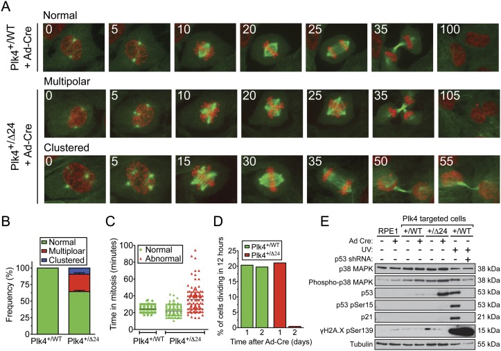
Figure 2.
Loss of Plk4 autoregulation leads to p53/p21 stabilization and cell cycle arrest. (A) Time-lapse images of cells stably expressing histone H2B-mRFP (red) and EYFP-α-Tubulin (green). Filming began 24 h after infection with Ad-Cre. Numbers in the top left refer to the time in minutes after nuclear envelope breakdown. (B) Quantification of the proportion of cells with normal, multipolar, or clustered spindle poles at the time of division. Cells were filmed for 16 h beginning 1 d after infection with Ad-Cre. Bars show the mean of >116 cells per condition from at least two independent experiments. Error bars represent the SEM. (C) Duration of mitosis in normal or abnormally dividing cells. Cells were filmed for 16 h beginning 1 d after infection with Ad-Cre. The line shows the mean of >116 cells per condition from at least two independent experiments. (D) Fraction of cells dividing in a 12-h period beginning 1 or 2 d after Ad-Cre infection. (E) Immunoblots of protein harvested 48 h after Ad-Cre infection or 12 h after UV irradiation.

