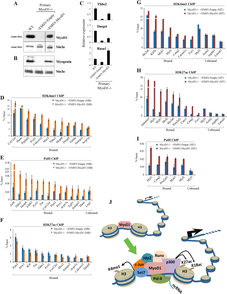
Figure 8.
Exogenous MyoD1 expression restores enhancer assembly in primary MyoD1−/− myoblasts. (A) Western blot detection of MyoD1 in nuclear extracts prepared from growing wild-type, MyoD1−/−, and MyoD1−/− (rescued) primary myoblasts that stably express exogenous MyoD1. Sin3A is shown as a loading control. (B) Western blotting of nuclear extracts of confluent myoblasts indicates that myogenin expression is restored to levels comparable with those of wild-type cells. (C) RT-qPCR analysis of three target genes associated with myoblast-specific enhancers bound by MyoD1. We note that each of the promoters associated with these genes was devoid of MyoD1 binding. (D–F) qChIP analysis was carried out to determine the impact of MyoD1 reconstitution in primary myoblasts on deposition of enhancer-related marks H3K4me1 (D), Pol II (E), and H3K27ac (F). (G–I) qChIP analysis for H3K4me1 (G), H3K27ac (H), and Pol II (I) was performed to examine indicated marks on myotube-specific enhancers after MyoD1 expression. Error bars for all qChIP and RT-qPCR data represent standard error of the mean (SEM). (J) Model in which MyoD1 acts to assemble enhancers in muscle. Interactions between MyoD1 and other factors are essential for enhancer assembly and acquisition of a transcriptionally active state. See the text for details.

