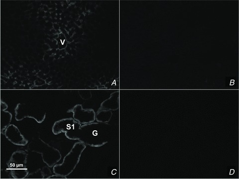
Figure 1. TAT1 detection by immunofluorescence microscopy in sections of liver (A and B) and kidney (C and D).

TAT1 is localized to the perivenous hepatocytes (A) and kidney proximal tubule cells (B) of wt tissues, whereas it is not detected in tat1−/− tissues (B and D). G, glomerulus; S1, proximal convoluted tubule segment S1; V, central vein.
