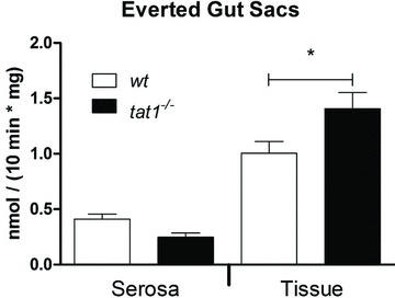
Figure 7. Significant accumulation of l-Phe in intestinal everted gut sacs.

Everted sacs of tat1−/− and wt mouse small intestine were incubated at 37°C for 10 min in Krebs buffer containing 100 μm l-Phe and l-[3H]Phe tracer. Radioactivity was counted inside of the sacs (serosa) and in the tissue (epithelial cells). Transport rates were calculated and normalized to weight of tissue. Represented are means ± SEM (n≥ 8). *P < 0.05 by unpaired Student's t test.
