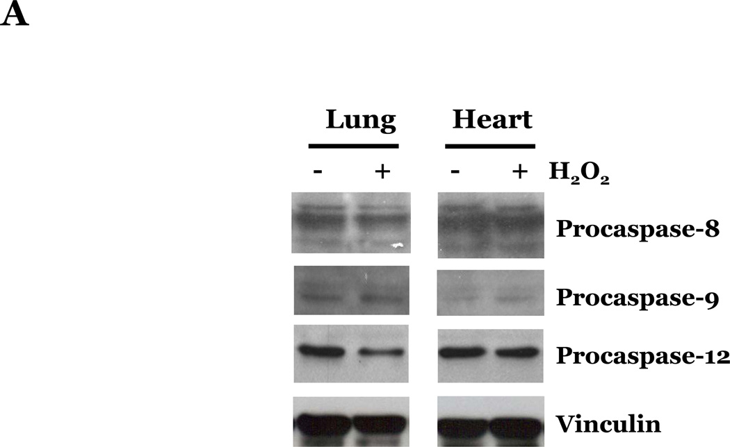
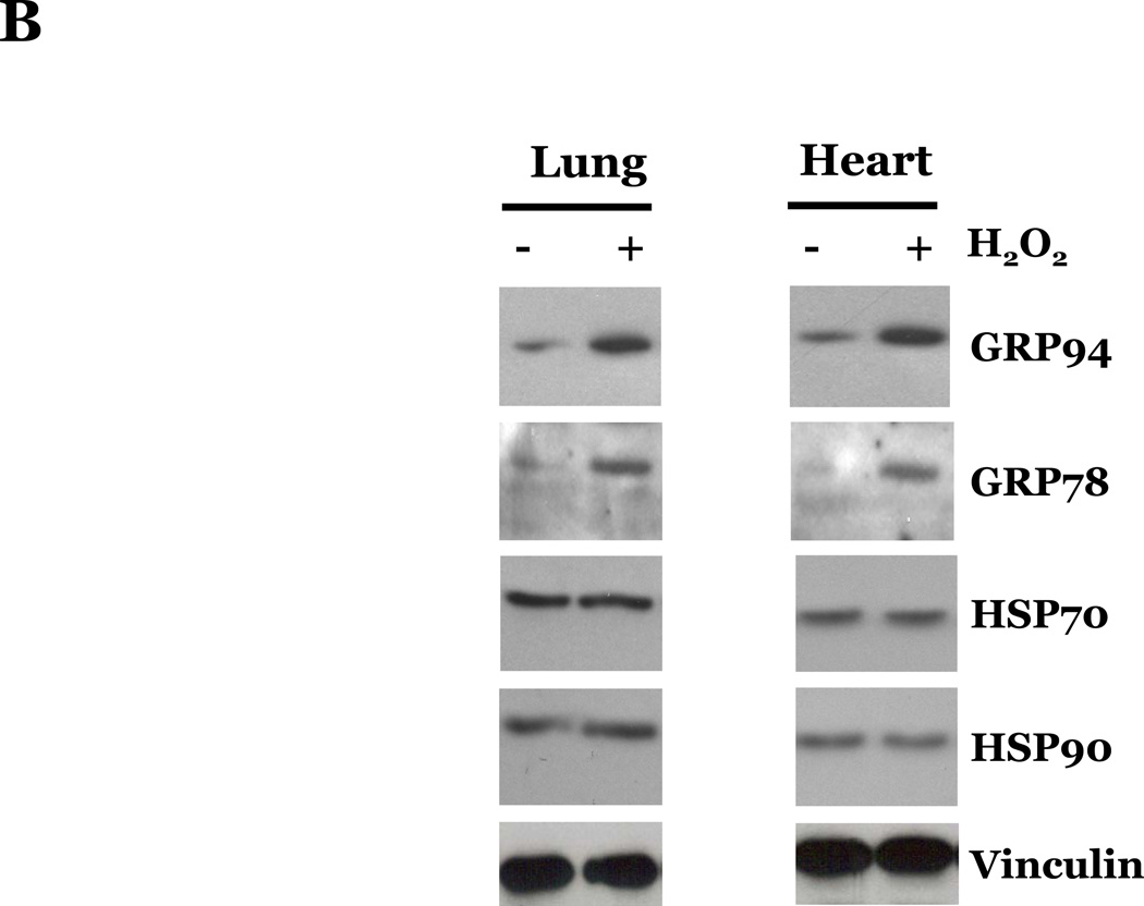
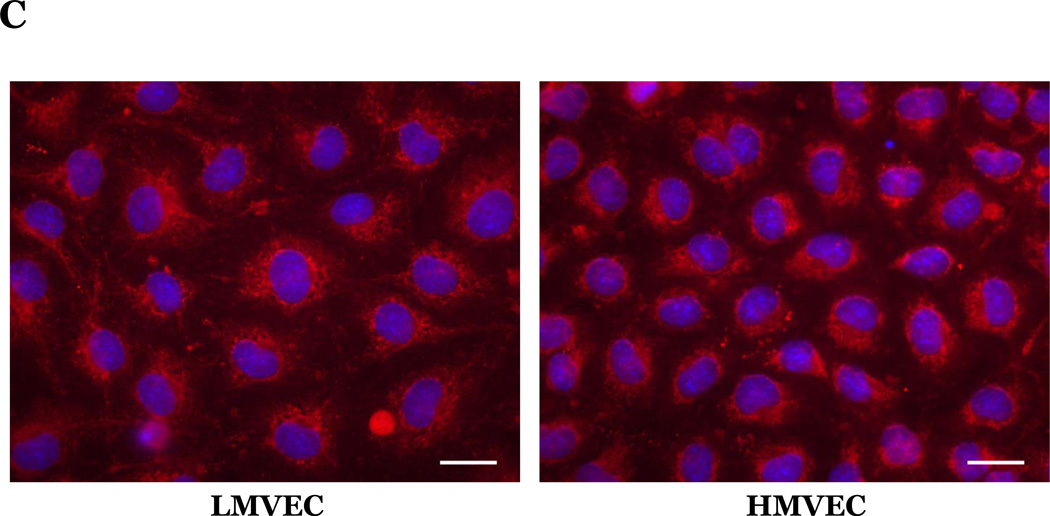
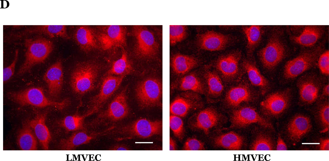
Figure 4. Oxidative stress promotes similar effects on caspase activation, UPR-associated chaperone proteins, and cytochrome c release in lung and heart microvascular endothelial cells.
Microvascular endothelial cells cultured in reduced serum medium were exposed to vehicle or H2O2 overnight. Equivalent amounts of lysates were resolved by SDS-PAGE and immunoblotted for indicated proteins. Representative gels are presented. n = 3. (panels A and B). Microvascular endothelial cells were cultured in reduced serum medium in settings of normoxia (panel C) or hyperoxia (panel D) for 24h. Cells were fixed and immunofluorescently stained for cytochrome c and viewed at 1000X magnification. Nuclei are counterstained with DAPI. Scale bar = 20µm. Representative images are presented. n = 3.


