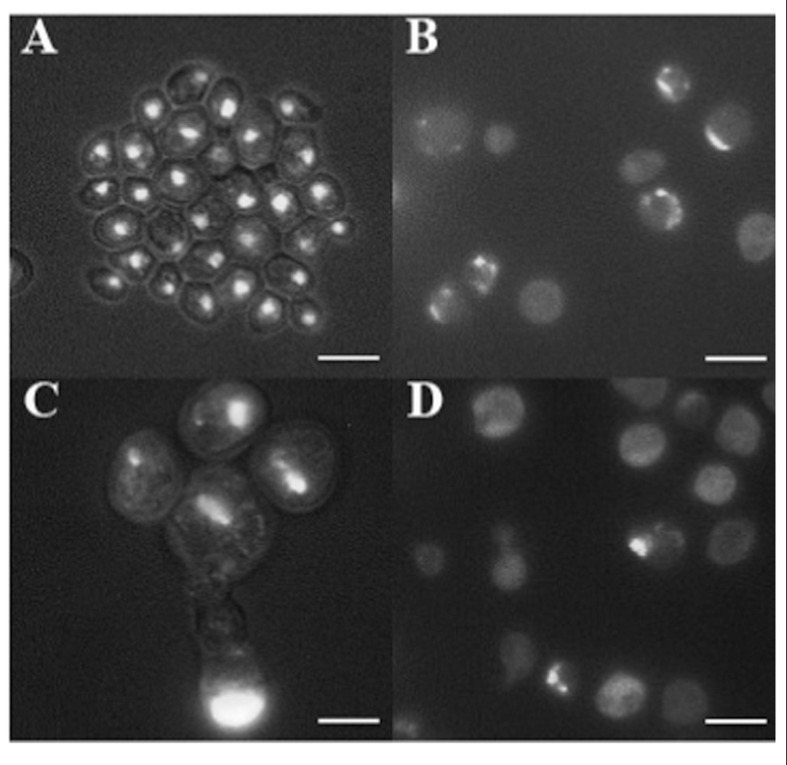
FIGURE 3.
DAPI staining of cells picked after 100 days from the smooth layer of colonies (A) and from three warts (B–D). Cells were fixed with 70% ethanol and stained with DAPI at the concentration of 1 μg/ml, and observed by fluorescence microscopy. Cells are shown at the same magnification. Bar, 10 μm.

