
Figure 1.
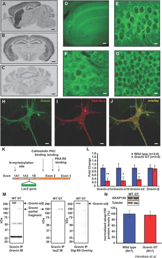
Expression of the α-isoform of gravin is reduced in gravin GT mice. A, A sagittal brain section showing gravin mRNA expression in the hippocampus, striatum, cortex, and olfactory bulb. B, A coronal brain section showing gravin mRNA in the hippocampus, cortex, amygdala, and striatum. C, Gravin mRNA is expressed at lower levels in the granular cell layer of the cerebellum. Scale bar, 800 μm. D, Gravin protein expression in the dorsal hippocampus. Scale bar, 200 μm. Higher magnification images of gravin protein expression in CA1 pyramidal cells (E), CA3 pyramidal cells (F), and granular cells of the dentate gyrus (G). Scale bar, 20 μm. Gravin (H) and PKA RII-α (I) are strongly colocalized (J) in cultured mouse hippocampal neurons. Scale bar, 20 μm. K, A schematic representation of the gravin gene. The gravin-α transcript consists of exons 1A1, 1A2, 2, and 3. The gravin-β transcript consists of exons 1B, 2, and 3. The gravin GT mice carry a LacZ insertion in the intron between exons 1A2 and 1B (Camus et al., 2001). This insertion site precedes the promoter region for the β transcript (Camus et al., 2001). L, Quantitative PCR experiments reveal reduced expression levels of gravin-α in gravin GT mice. mRNA levels for gravin-α1a (Student's t test, n = 6, p = 0.009), gravin-α1b (Student's t test, n = 3, p = 0.039), and gravin-α2 (Student's t test, n = 6, p = 0.014). Gravin-β expression was not affected in gravin GT mice (Student's t test, n = 6, p = 0.444). M, Gravin GT mice express a truncated form of gravin-α of ∼150 kDa (left panel), which contains LacZ (middle panel) but lacks an RII binding site (right panel). N, Hippocampal AKAP150 protein levels are not altered in gravin GT mice (n = 7 for both groups; Student's t tests, p = 0.79). *p < 0.05; **p < 0.01. Error bars indicate SEM.