
Figure 4. Transdifferentiation of c-kit–positive HSCs.
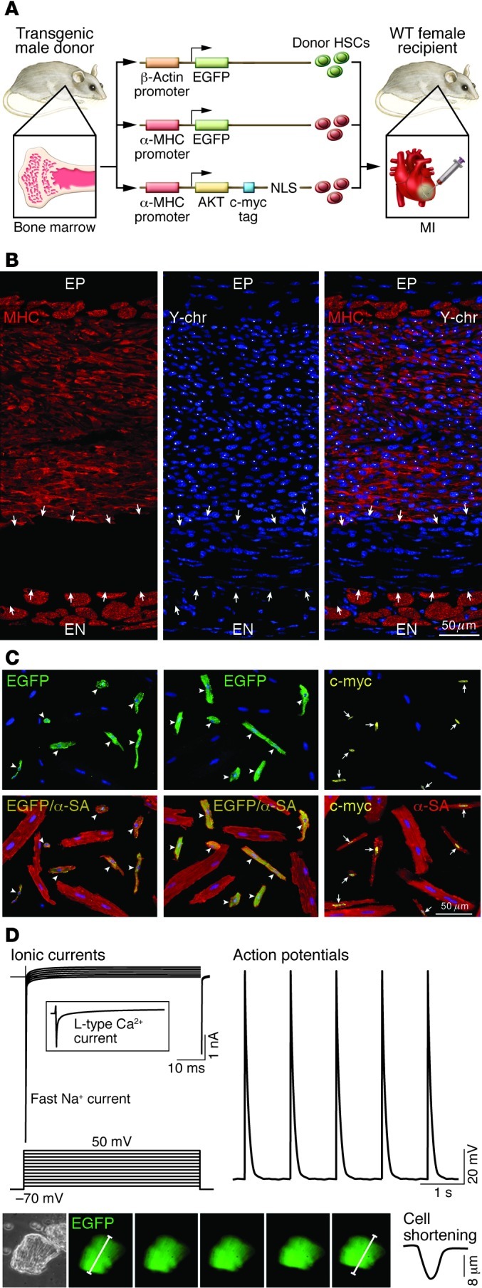
(A) Schematic representation of transgene constructs used in the generation of donor mice for the acquisition of HSCs to be delivered after infarction. In each case, the promoter that controls the ubiquitous (β-actin) or myocyte-restricted (α-MHC) expression of the transgene (EGFP or c-myc–tagged nuclear-targeted Akt) is shown. c-kit–positive HSCs from donor males were injected intramyocardially in wild-type female infarcted mice. NLS, nuclear localization signal (ref. 75). (B) Infarcted female mouse treated with male HSCs. Regenerated myocytes (left; MHC, red) carry the Y chromosome (center; Y-chr; white dots in nuclei). Merged image is shown at right. Arrows indicate non-regenerated infarct. A thin layer of spared myocytes is present in the epimyocardium (EP) and endomyocardium (EN) (ref. 75). Scale bar: 50 μm. (C) Examples of myocytes isolated from the regenerated myocardium of mice injected with HSCs collected from β-actin EGFP, α-MHC EGFP, or α-MHC c-myc–tagged nuclear-targeted Akt mice. Top panels illustrate the localization of EGFP (left and middle, green; arrowheads) and c-myc (right, yellow nuclei; arrows) in newly formed cardiomyocytes. Bottom panels show the colocalization of α-SA and EGFP (yellow, arrowheads) and of α-SA and c-myc (yellow nuclei, arrows). Large spared myocytes negative for EGFP or c-myc are also present (ref. 75). Scale bar: 50 μm. (D) Myocyte-specific ionic currents and action potentials of a cell isolated from the regenerated infarcted myocardium. EGFP-positive myocytes show contractile activity (bottom). Original magnification, ×200. Reproduced with permission from Proceedings of the National Academy of Sciences of the United States of America (ref. 75; copyright 2007, National Academy of Sciences, USA).