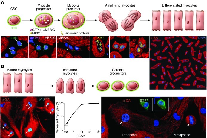
Figure 5. Cellular mechanisms of myocardial homeostasis.
(A) Lineage relationship between CSCs and cardiomyocytes: CSC commitment results progressively in the formation of myocyte progenitors, myocyte precursors, and transit-amplifying myocytes, which divide and differentiate into mature myocytes. CSCs are c-kit–positive lineage-negative cells. Progenitors express c-kit and myocyte-specific transcription factors (GATA4, Nkx2.5, MEF2C) but do not show sarcomeric proteins. Precursors possess c-kit, myocyte-specific transcription factors, and sarcomeric proteins. Amplifying cells have myocyte nuclear and cytoplasmic proteins but are c-kit negative. This hierarchy is shown by immunolabeling and confocal microscopy (bottom). Micrographs from left to right show lineage-negative CSCs (arrows); myocyte progenitors (arrowheads) expressing c-kit, GATA4, and MEF2C; myocyte precursor (asterisk) showing c-kit, MEF2C, and α-SA; and three transit-amplifying myocytes (α-SA). Chromosomes are organized in anaphase-telophase and are Ki67 positive. Original magnification, ×700. Terminally differentiated cardiomyocytes from the human heart are shown at far right. Original magnification, ×100. Reproduced with permission from Proceedings of the National Academy of Sciences of the United States of America (ref. 9; copyright 2003, National Academy of Sciences, USA). (B) The postulated process of dedifferentiation of postmitotic myocytes to immature myocytes and cardiac progenitors is represented schematically. Micrographs document that culture of differentiated myocytes is not coupled with reentry into the cell cycle and reversal to a primitive state. From left to right, adult cardiomyocytes in vitro show disassembly of the contractile apparatus and express the senescence-associated protein p16INK4a (bright blue, arrowheads; original magnification, ×250). The percentage of senescent myocytes increases with time. Fetal myocytes, isolated from a reporter mouse in which EGFP is driven by the c-kit promoter, divide in vitro (arrows) but do not express c-kit. Original magnification, ×500. Insets show c-kit–positive EGFP-positive CSCs used as a positive control. Reproduced with permission from Circulation Research (ref. 61).

