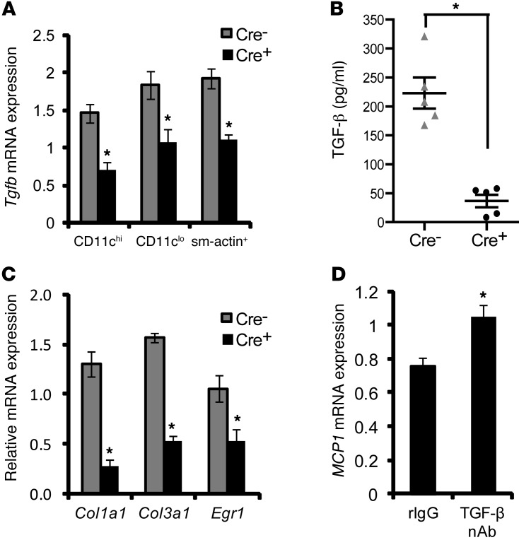
Figure 6. Evidence of a link between decreased TGF-β and increased MCP1 in lesions of WD-fed Ldlr–/– mice transplanted with bone marrow from Cre+ mice.
(A) LCM RT-qPCR analysis of Tgfb mRNA in RNA obtained from CD11chi, CD11clo, and sm-actin+ regions of atherosclerotic lesions of Cre– and Cre+ mice (n = 5 mice per group). (B) ELISA-based measurement of latent TGF-β from extracts of atherosclerotic lesions of Cre– and Cre+ mice (n = 5 mice per group). (C) Col1a1, Col3a1, and Egr1 mRNA in RNA obtained from sm-actin+ regions of atherosclerotic lesions of Cre– and Cre+ mice (n = 5 mice per group). (D) Analysis of MCP1 mRNA expression in RNA obtained from the intima of atherosclerotic lesions of 10-week WD-fed Ldlr–/– mice treated with rat IgG (rIgG) or a neutralizing rat IgG antibody against TGF-β (TGF-β nAb) (100 μg per mouse) on days 1, 3, and 8 prior to sacrifice. The data were normalized to expression of Gapdh (n = 6 mice per group). For all panels, *P < 0.05. Symbols represent individual mice; horizontal bars indicate the mean.

