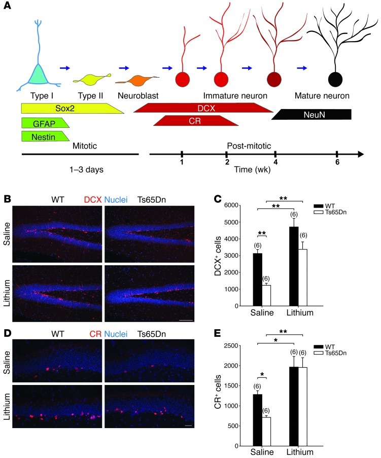
Figure 1. Lithium restored the number of newborn neurons in DG of adult Ts65Dn mice.
(A) Expression of molecular markers during adult DG neurogenesis. During the mitotic phase, 2 types of progenitors proliferate in the SGZ of the DG: RGL progenitors (type I cells, expressing Nestin, GFAP, and Sox2) and amplifying progenitors (type II cells, expressing Sox2 only). Before becoming postmitotic, the progenitors undergo a short intermediate stage (neuroblasts), commit to the neuronal fate, and begin expressing DCX. In the postmitotic phase, DCX-positive newborn neurons derived from neuroblasts undergo a morphological and physiological maturing process with the sequential expression of CR and NeuN upon final maturation. (B–E) Five- to six-month-old Ts65Dn and WT mice were treated with lithium or saline for 4 weeks, and the number of newborn neurons was evaluated through immunohistochemistry for DCX or CR (red) and counterstained with Hoechst-33342 (blue). (B) Immunoreactivity and (C) number of DCX+ newborn neurons in saline- or lithium-treated Ts65Dn and WT mice. 2-way ANOVA: genotype (F1,20 = 19.285, P < 0.001), treatment (F1,20 = 25.570, P < 0.001), genotype × treatment (F1,20 = 0.584, P = 0.454). (D) Immunoreactivity and (E) number of CR+ newborn neurons in DG of saline- or lithium-treated Ts65Dn and WT mice. 2-way ANOVA: genotype (F1,20 = 2.413, P = 0.136), treatment (F1,20 = 26.602, P < 0.001), genotype × treatment (F1,20 = 2.247, P = 0.149). Numbers in parentheses indicate the number of animals in each group. *P < 0.05; **P < 0.01, Tukey’s post hoc test. Scale bars: 100 μm (B); 25 μm (D).

