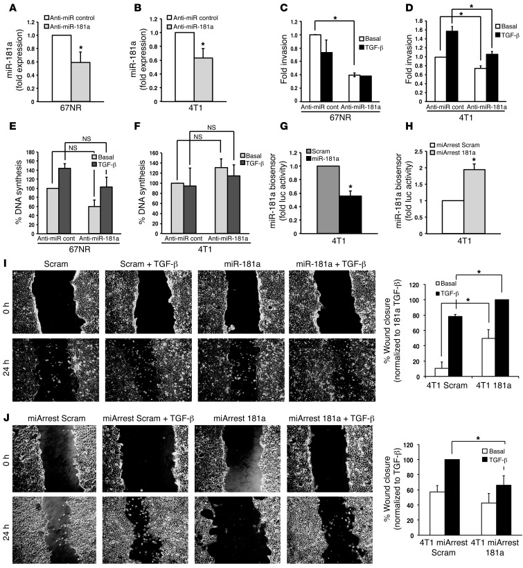
Figure 3. Inhibition of miR-181a attenuates TGF-β–mediated EMT, invasion, and migration.
(A and B) An antagomir against miR-181a (anti–miR-181a) was transiently transfected into 67NR or 4T1 cells, resulting in decreased expression of miR-181a, as determined by semiquantitative real-time PCR. (C and D) The invasiveness of miR-181a–manipulated 67NR and 4T1 cells in response to TGF-β1 (5 ng/ml) treatment was significantly reduced by miR-181a inactivation. (E and F) The proliferation of miR-181a–manipulated 67NR and 4T1 cells in response to TGF-β1 (5 ng/ml) treatment was unaffected by miR-181a inactivation. (G and H) 4T1 cells engineered to stably express miR-181a (G) or a miR-181a sponge (H) were transiently transfected with a renilla luciferase miR-181a biosensor and CMV–β-gal, which was used to control for differences in transfection efficiency. miR-181a was shown to significantly elevate miR-181a activity, while miR-181a antagonists were shown to significantly reduce miR-181a activity. (I and J) The ability of TGF-β1 (5 ng/ml) to induce the migration of 4T1 cells with elevated (I) or reduced (J) miR-181a activity was significantly stimulated by miR-181a activation (I) or was significantly inhibited by miR-181a inactivation (J). Original magnification, ×20. All data are the mean ± SEM. n = 3. *P < 0.05, Student’s t test.

