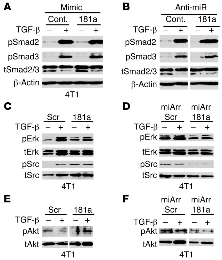
Figure 4. miR-181a expression enhances Erk1/2, Akt, and Src signaling in breast cancer cells.
(A–F) 4T1 cells that harbored manipulated miR-181a activity as indicated were stimulated with TGF-β1 (5 ng/ml) for 30 minutes. Afterward, the phosphorylation and expression of Smad2/3, Erk1/2, Akt, and Src were measured by immunoblotting as indicated. Shown are representative images from 3 independent experiments. Scr, scrambled control vector; miArr, miArrest vector. Lanes in B, E, and F were run on the same gel, but were noncontiguous (white lines).

