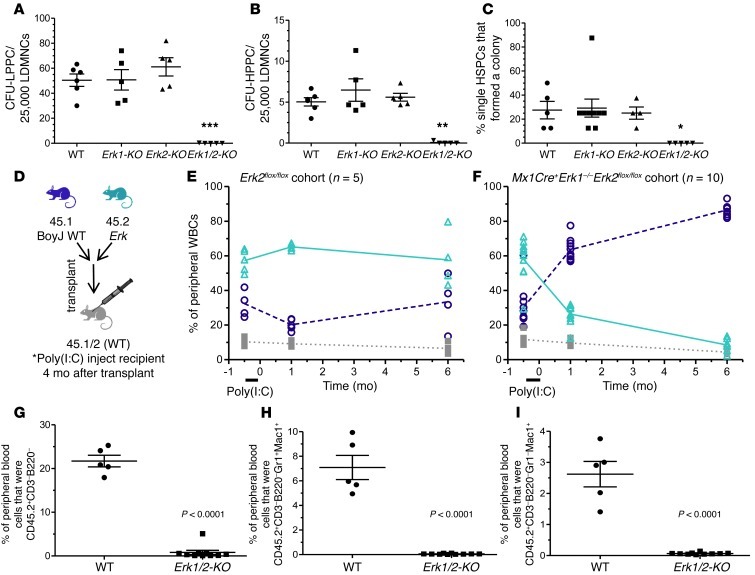
Figure 2. Granulocytopoiesis and monocytopoiesis require Erk.
(A and B) Erk1/2-KO cells form few colonies in LPPC-HPPC assays. **P < 0.01; ***P < 0.001, Erk1 /2-KO vs. all, 1-way ANOVA with Bonferroni’s correction. (C) Single Erk1/2-KO SLAM-LS cells fail to produce colonies and show no evidence of expansion using Hoechst-based cell detection (not shown). *P < 0.05 Erk1/2-KO vs. all, 1-way ANOVA with Bonferroni’s correction. (D) Lethally irradiated CD45.1/2+ mice received mixed CD45.2+ (Erk mutant) and CD45.1+ (WT) marrow cells at a one-to-one ratio. (E and F) After Cre induction (4 months following transplantation), the WT CD45.2+ recipients demonstrated stable chimerism, but the Erk1/2-KO recipients experienced rapid and progressive loss of CD45.2+ cells, as measured in the peripheral blood (for WT vs. Erk1/2-KO 45.2+ chimerism, data not significant at –0.5 months, P < 0.001 at 1 and 6 months, 2-way ANOVA with Bonferroni’s correction). (G–I) Peripheral blood flow cytometric analyses after Cre induction demonstrate ablated Erk1/2-KO-derived CD45.2+ circulating nonlymphoid cells (CD3–B220–), granulocytes (CD3–B220–Gr1+Mac1+), and monocytes (CD3–B220–Gr1–Mac1+). P < 0.0001, WT vs. Erk1/2-KO, Student’s 2-tailed unpaired t test.

