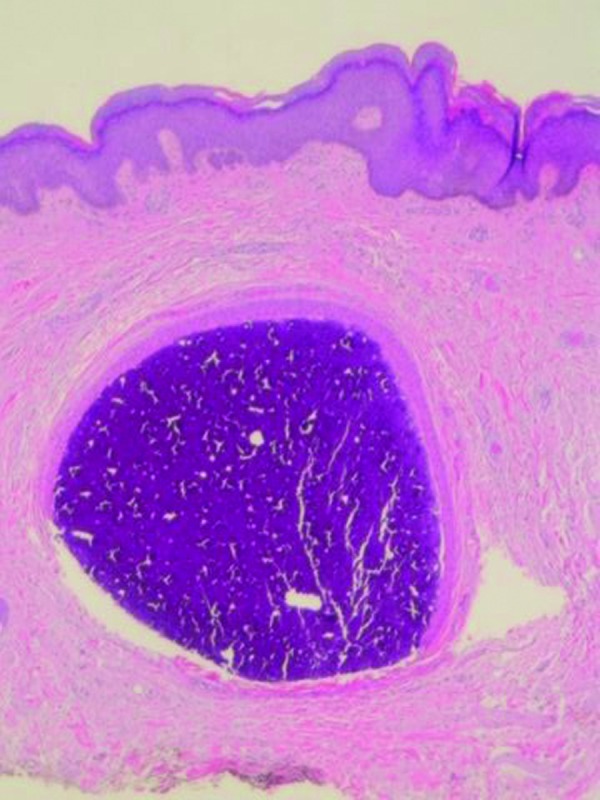
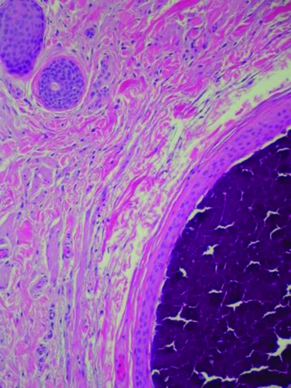
Figures 5A and 5B.


Microscopic examination—lower (left) and higher (right) magnification—of scrotal nodule shows basophilic-staining calcified mass surrounded by an epithelial lining in the dermis; eccrine structures, composed of similar appearing cells as the epithelial lining surrounding the amorphous mass of calcium, are present in the adjacent dermis (hematoxylin and eosin: X4; X10).
