
Fig. 1.
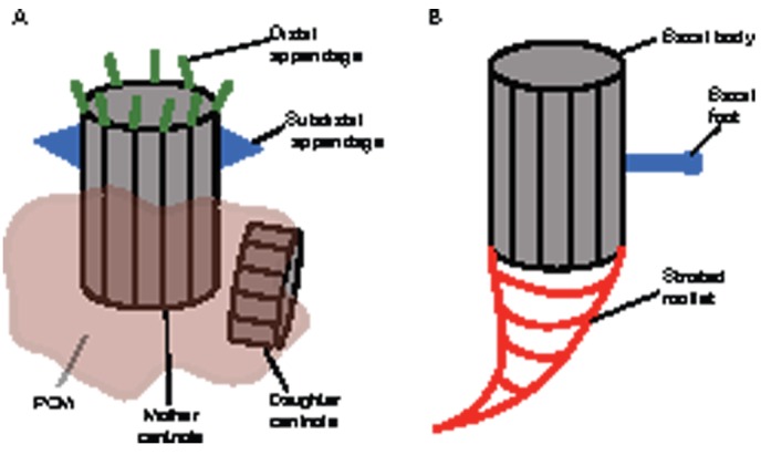
The anatomy of vertebrate centrosomes and centrioles. (A) Schematic diagram of typical centrosome and centriole organization in most vertebrate cells. Centrosomes are composed of two orthogonally oriented centrioles that are surrounded by PCM. The mother centriole is distinguished by two sets of appendages, the subdistal and distal appendages (Paintrand et al., 1992), which are thought to be required for anchoring microtubules at the centriole or forming transitional fibers that contact the cell cortex, respectively (Dawe et al., 2007). (B) Organization of the basal body (centriole) in a cell from a multiciliated epithelium. The proximal side of the basal body is associated with a matrix, which extends into specific and striated structures called rootlets (Klotz et al., 1986). PCM, pericentriolar material.