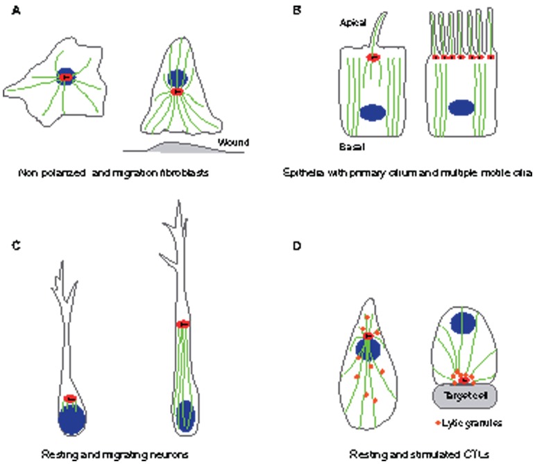
Fig. 2.
Examples of centrosome positioning that depend on cell type and cell state. (A) Fibroblasts: in non-polarized fibroblasts (left), the centrosome is located near the center of the cell and is physically linked to the nucleus, with microtubules radiating from the centrosome to the cell cortex. During wound healing (right), the fibroblast centrosome often becomes oriented between the nucleus and the leading edge. (B) Epithelial cells: in polarized epithelial cells (left), centrosomes are located on the apical surface of the cell. The apical localization of the centrosome is accompanied by a loss of radial microtubule organization and the formation of a predominantly apical–basal array of microtubules. The mother centriole of the centrosome becomes a basal body, which gives rise to a primary cilium. In multiciliated epithelial cells (right), hundreds of centrioles are assembled at once in a single cell, leading to the formation of multiple cilia. (C) Neurons: in resting neurons (left), the centrosome is found in close proximity to the neurite that becomes the axon. In migrating neurons (right), the centrosome is positioned ahead of the nucleus, with microtubules connecting the centrosome and the nucleus. (D) Lymphocytes: in cytotoxic T lymphocytes (CTLs) that are not interacting with a target (left), the centrosome is located near the nucleus, and lytic granules are distributed all along the microtubules. After the CTL is stimulated (right), the centrosome directs the delivery of lytic granules by moving along microtubules to the plasma membrane and then to the point of secretion for releasing lytic granules to the immunological synapse. Red ovals indicate the centrosome, with centrioles indicated as black lines within. Blue ovals indicate nuclei and green lines indicate microtubules.

