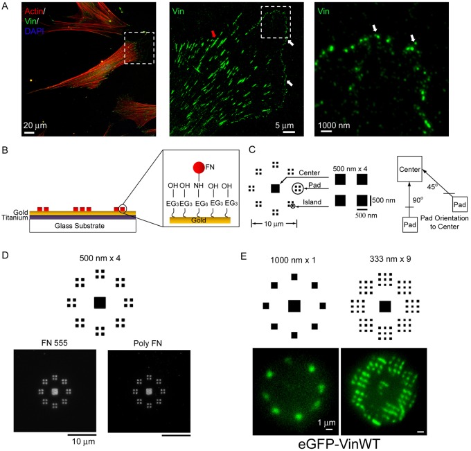
Fig. 1.
Cells adhere to nanopatterned FN and form adhesive structures. (A) Nanoscale nascent adhesions (vinculin) formed at the lamellipodium and leading edge of a fibroblast (mature adhesions: red arrowhead; nascent adhesions: white arrowhead). (B) FN patterns on PDMS stamps are transferred onto the substrates with NHS esters resulting in the transfer and tethering of FN molecules onto the substrate. (C) Adhesive zone of nanoislands (500 nm×4 shown); each zone consists of a center square (2×2 µm) surrounded by eight radially distributed adhesion pads. Each adhesion pad consists of 1, 2, 4 or 9 square nanoislands. Adhesive pads are presented in two orientations (45°, 90°) relative to the center pad. (D) FN nanopatterns retain high spatial fidelity in culture as visualized by direct imaging of Alexa-Fluor-555-labeled FN (FN 555) or indirect immunostaining using a polyclonal antibody against FN (poly FN). (E) Cells expressing GFP–vinculin assemble vinculin-containing FAs on nanopatterns.

