Fig. 2.
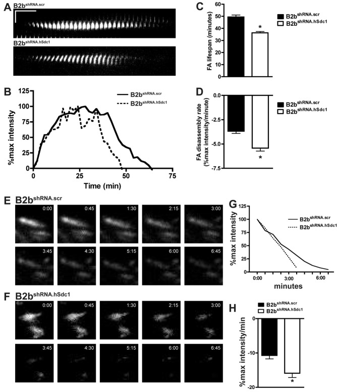
Syndecan-1 slows FA disassembly in lung epithelial cells. (A–D) Migrating B2bshRNA.scr and B2bshRNA.hSdc1 cells stably expressing paxillin–eGFP were observed by time-lapse TIRF microscopy (supplementary material Movie 1). (A) Kymographs revealed a longer FA lifespan in B2bshRNA.scr compared to B2bshRNA.hSdc1 cells. Horizontal line: 10 min; vertical line: 5 µm. (B) Normalized fluorescent intensity (% of max intensity) of the FAs in panel A over time. (C) The total FA lifespan and (D) the rate of the FA disassembly were determined for migrating lung epithelial cells. *P<0.0001 by Student's t-test; n = 4 independent experiments. (E–H) B2bshRNA.scr and B2bshRNA.hSdc1 stably expressing paxillin–eGFP were treated with nocodazole (10 µM; 4 h). After washing the cells with fresh medium, FA disassembly was observed with time-lapse TIRF microscopy in (E) B2bshRNA.scr and (F) B2bshRNA.hSdc1 cells. (G) The intensity curves of the FAs in panels E and F. (H) The FA disassembly rate was determined in B2bshRNA.scr and B2bshRNA.hSdc1 cells. *P<0.05 by Student's t-test; n = 3 independent experiments.

