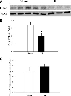
Fig. 7.
(A) Mesenteric artery homogenates from sham and IH group rats were immunoprecipitated with PKCδ (78 kDa) antibody and probed for PDK-1 (58–68 kDa). There was diminished interaction between PDK-1 and PKCδ in arteries from rats in the IH group compared with those from the sham group (B). Total PKCδ levels were normalized to Coomassie Blue staining and were not affected by exposure to IH (C). *P < 0.05 versus sham. The images shown include tissue from four rats per group and are representative of three replicates. A.U., arbitrary units.

