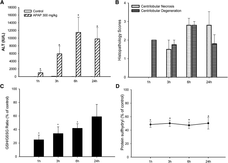
Fig. 1.
Time course for the effect of APAP on serum ALT, histopathology score, hepatic GSH/GSSG ratio, and protein sulfhydryl depletion (as measured by SDS-PAGE). APAP (300 mg/kg) was administered by oral gavage, then the blood and liver samples were collected at the indicated times. (A) APAP statistically significantly increased serum ALT values, indicative of hepatocellular injury. Liver histopathology lesions were scored on a 5-point scale. (B) All time-matched control animals had scores of zero. (C) Hepatic GSH and GSSG levels were measured by UPLC-MS, and the GSH/GSSG ratio was used as an indicator of oxidative stress. The GSH/GSSG ratio was statistically significantly decreased compared with control levels from 1 to 6 hours. The GSH/GSSG ratio was still decreased at 24 hours, but the decrease was not statistically significant. (D) Protein thiols were labeled with maleimide-IRDye, scanned, and quantified with a near-infrared scanner. The same samples were resolved on a duplicate gel followed by Coomassie Blue staining, and the protein sulfhydryl levels were normalized to the Coomassie Blue staining. APAP at 300 mg/kg produced a profound decrease of protein thiols at 1 hour through 24 hours. *ALT, GSH/GSSG ratio, and protein thiols of 300 mg/kg group were statistically significantly decreased (P < 0.05) compared with the control group. Histopathology scores were not statistically analyzed. Values are mean ± S.E.M.

