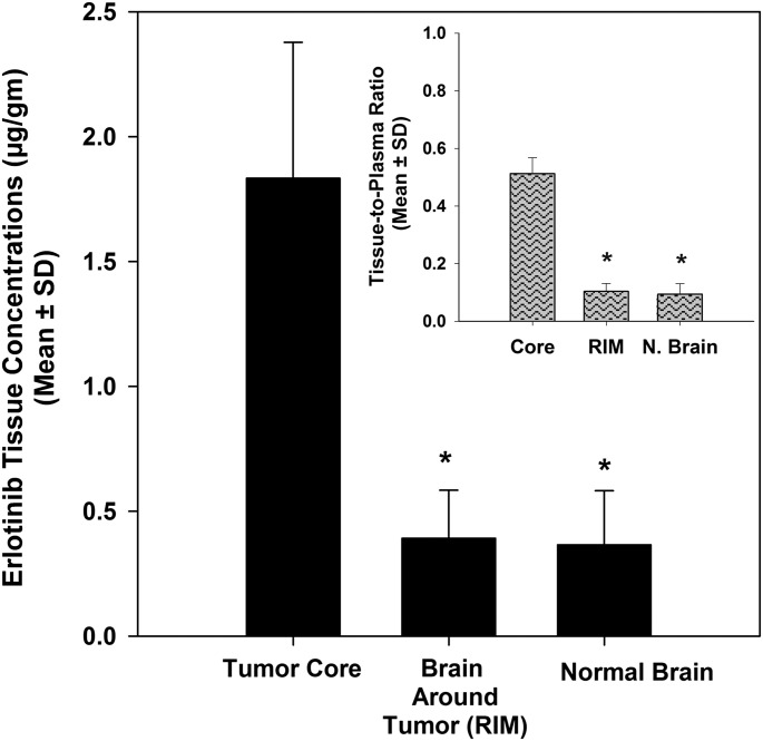
Fig. 3.
Regional distribution of erlotinib in the U87 rat xenograft model. Erlotinib concentrations in the tumor core were significantly greater than those in the brain around the tumor and normal brain (contralateral hemisphere). The corresponding brain-to-plasma ratios (inset) suggests that the BBB may be intact in areas away from the tumor, indicated by the restricted delivery of erlotinib to the brain around the tumor and the normal contralateral hemisphere. The values are presented as the mean ± S.D. *P < 0.05, compared with tumor core; n = 6.

