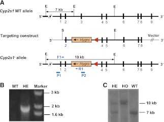
Fig. 1.
Targeted disruption of the mouse Cyp2s1 gene. (A) targeting strategy for Cyp2s1. The Cyp2s1 gene sequence is indicated by a thick line, whereas sequences from cloning vectors are shown in thin lines. Cyp2s1 exons are indicated as solid boxes. The positions of selected restriction sites (E, EcoR V; S, Spe I) are indicated. PCR primers used for genotyping (F1/R1) are shown as small thin arrows. DNA probes used for Southern blot analysis, P1 (external probe, 1.3-kb in size) and P2 (internal probe, 1.3 kb in size), are shown as blue bars. The diagnostic EcoR V fragments detected by probe P1 in WT allele (7 kb) and in targeted (Cyp2s1−) allele (10 kb), as well as the positions of PCR primers used for detecting Cyp2s1− allele, are indicated. (B) PCR analysis of genomic DNA from WT and Cyp2s1+/− mice. The expected 1.9-kb product was detected in the heterozygotes. Selected bands of a 1-kb DNA size marker are shown. (C) Southern blot analysis of genomic DNA with the external probe P1. Expected EcoR V fragments were detected in heterozygous (HE), homozygous (HO), and WT mice.

