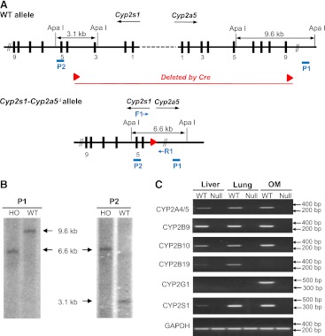
Fig. 3.
Confirmation of gene cluster deletion in the Cyp2a(4/5)bgs-null (Cyp2s1-Cyp2a5Δ/Δ) mice. (A) strategy of Southern blot analysis. DNA probes P1 and P2 are shown as blue bars. Genomic DNA was digested by Apa I. Probe P1 would detect a 9.6-kb fragment from Cyp2a5 WT allele, whereas probe P2 would detect a 3.1-kb fragment from Cyp2s1 WT allele; both probes would detect a 6.6-kb fragment from the Cyp2s1-Cyp2a5Δ allele. (B) Southern blot analysis of WT and Cyp2s1-Cyp2a5Δ/Δ (HO) mice. Ten micrograms of genomic DNA were used for each lane. (C) RNA-PCR analysis of CYP2A4/5, CYP2B9/10/19, CYP2G1, and CYP2S1 expression in liver, lung, and OM of WT and Cyp2a(4/5)bgs-null mice. Tissues from 2-month-old (one male and one female) mice were pooled for total RNA preparation. PCR products were analyzed on a 1.5% agarose gel, and visualized by staining with ethidium bromide. The positions of selected fragments of a 100-bp DNA marker are indicated.

