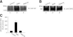
Fig. 3.
PKC specificity of OAT1 ubiquitination in the rat kidney. (A) rat kidney slices were treated with PKC-activator PMA (1 μM) in the presence or absence of the PKC-inhibitor staurosporine (St, 2 μM) for 30 minutes. The treated slices were then lysed, and OAT1 was immunoprecipitated with anti-OAT1 antibody, followed by immunoblotting (Ib) with anti-ubiquitin antibody P4D1. (B) the same immunoblot as seen in (A) was reprobed by anti-OAT1 antibody. (C) densitometry plot of results from (A) as well as from other experiments. Values are mean ± S.E. (n = 3).

