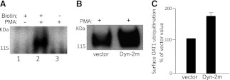
Fig. 5.
Cell-surface OAT1 ubiquitination. (A) cell-surface proteins of OAT1-expressing cells were labeled with or without the membrane-impermeable reagent biotin. The cells were treated with or without PMA for 30 minutes. The labeled cells were lysed. OAT1 was then immunoprecipitated with subsequent streptavidin pull-down to isolate cell-surface OAT1, followed by immunoblotting with anti-ubiquitin antibody. (B) OAT1-expressing cells were transfected with the cDNA encoding dominant negative mutant of dynamin-2 (Dyn-2m, empty vector as control). The transfected cells were treated with PMA (1 μM) for 30 minutes. Cell-surface OAT1 was isolated by cell-surface biotinylation, as described in (A), followed by immunoblotting with anti-ubiquitin antibody. (C) densitometry plot of results from (B) as well as from other experiments. Surface OAT1 in PMA-treated cells was expressed as the percentage of surface OAT1 in control cells. The values are mean ± S.E. (n = 3).

