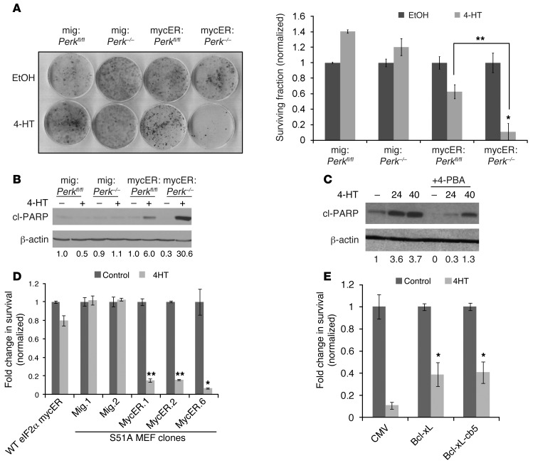
Figure 3. Loss of UPR signaling results in c-Myc–induced caspase-dependent apoptosis.
(A) Perkfl/fl or Perk–/– MEFs infected with control (mig) or mycER–expressing retroviruses were treated with EtOH or 4-HT, and clonogenic survival was assayed (left panel); colonies were counted, and the surviving fraction is presented (right panel) normalized to each untreated control (error bars represent SEM; n = 3; **P < 0.0001, *P < 0.0003). (B) MEFs were treated with 4-HT for 24 hours, followed by immunoblotting for cleaved PARP (c-PARP). (C) mycER:Perk–/– MEFs were treated with 4-HT in the presence or absence of 4-PBA (5 mM), and immunoblotting was performed for cleaved-PARP. (D) S51A-eIF2α knock-in MEFs infected with control (mig) or mycER-expressing retroviruses were treated with ethanol or 4-HT, and survival was assayed (compared with WT MEFs expressing mycER; **P < 0.00002, *P < 0.002, Student’s 2-tailed t test). (E) mycER:Perk–/– MEFs were transfected with CMV control or Bcl-xL–expressing plasmids (WT or ER-targeted cb5), treated with 4-HT, cultured for 72 hours, and stained with crystal violet, and cell survival was quantified (*P < 0.001, Student’s 2-tailed t test).

