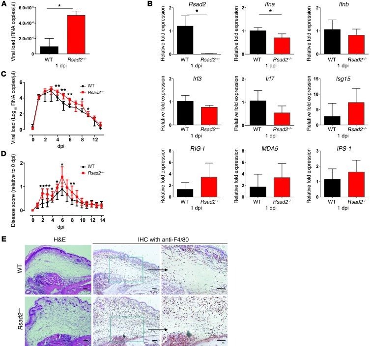
Figure 8. Viperin modulates CHIKV replication and disease pathology in mice.
(A) WT and Rsad2–/– mice (n = 4–6 per group) were infected with 106 PFU CHIKV in the footpad. Viral RNA was isolated from the footpads a 1 dpi and quantified by qRT-PCR against the negative-strand nsP1 RNA. *P < 0.05, Mann-Whitney U test. (B) qRT-PCR was used to determine the expression of the indicated genes in infected footpads of mice. Data (mean ± SD) were normalized to GAPDH and shown as fold expression relative to WT. *P < 0.05, Mann-Whitney U test. (C) WT and Rsad2–/– mice (n = 6 per group) were infected with 106 PFU CHIKV in the footpad. Blood was collected daily at 0–14 dpi for quantification of viral load as described in A (mean ± SD). *P < 0.05, **P < 0.01, Mann-Whitney U test. (D) Quantification of joint swelling in WT and Rsad2–/– mice. Size of infected joint was measured daily for 14 days and expressed as disease score relative to day 0 (preinfection). Data (mean ± SD) are representative of 2 independent experiments. *P < 0.05, **P < 0.01, Mann-Whitney U test. (E) Histology of CHIKV-induced inflammation in footpads of WT and Rsad2–/– mice was analyzed by H&E staining and IHC staining with anti-F4/80 antibody at 6 dpi. Boxed regions are shown at higher magnification at right. Scale bars: 100 μm.

