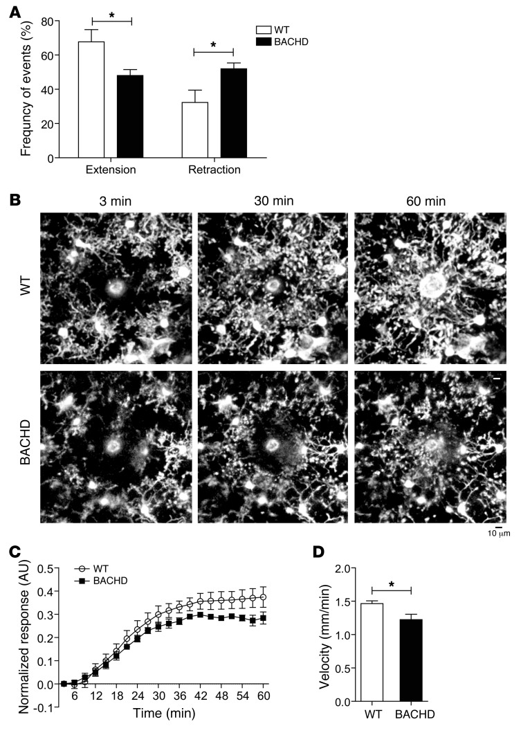
Figure 3. Microglia show defective basal process dynamics and delayed responses to focal laser ablation in the cortex of BACHD mice in vivo.
Microglia from WT;Cx3cr1GFP/+ (WT) or BACHDTg/+;Cx3cr1GFP/+ (BACHD) mice were imaged in vivo using 2-photon microscopy at 12–18 months of age. (A) Baseline time-lapse imaging of microglia demonstrated reduced extension and increased retraction of microglial processes over 10 minutes in BACHDTg/+;Cx3cr1GFP/+ compared with WT;Cx3cr1GFP/+ control mice. *P < 0.05 (t test). (B) Tissue ablation with a laser (white zone in center) results in rapid extension of microglial processes toward the site of injury in WT;Cx3cr1GFP/+ mice. In contrast, microglia from BACHDTg/+;Cx3cr1GFP/+ mice show a delayed response over a 60-minute period. (C) Quantification of process extension toward the site of laser ablation. P < 0.001 (2-way ANOVA). (D) The average velocity of process extension is significantly decreased in microglia from BACHDTg/+Cx3cr1GFP/+ mice over a 45-minute period. *P < 0.05 (t test). Values are mean ± SEM. n = 4–5 mice. Scale bar: 10 μm.

