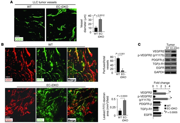
Figure 2. Enlarged leaky tumor vessels and elevated VEGFR2 signaling by loss of endothelial epsins 1 and 2.
(A) LLC tumor vessels in WT and EC-iDKO mice revealed by CD31 immunostaining. Quantification of vessel diameter is on right. (B) LLC tumor vessels in WT and EC-iDKO mice after FITC-dextran perfusion observed by CD31 immunostaining. Quantification of perfused vessels and leaked FITC-dextran area is on right. (C) Western blotting analysis of indicated proteins in tumor tissue lysates from WT and EC-iDKO mice. Quantification of fold change normalized according to loading control GAPDH is on right. n = 10 (A and B); n = 5 (C). Scale bars: 50 μm (A and B).

