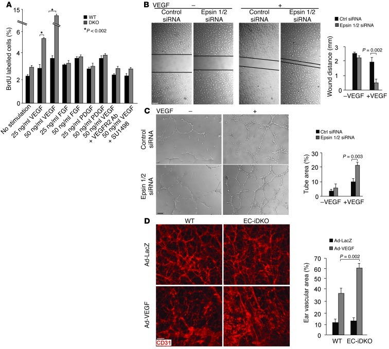
Figure 8. Loss of endothelial epsins 1 and 2 augments EC proliferation and migration and increases VEGF-induced ear neovascularization in mice.
(A) VEGF but not FGF or PDGF increased proliferation of DKO MECs relative to WT MECs measured by BrdU labeling. The increase is abrogated by inhibitors of VEGFR2. (B) HUVECs transfected with control or epsin 1 and 2 siRNAs were subjected to a monolayer “wound injury” assay in the absence or presence of VEGF-A (50 ng/ml) for 12 hours. (C) HUVECs transfected with control or epsin 1 and 2 siRNAs were cultured on Matrigel for 16 hours in the absence of presence of VEGF-A (50 ng/ml). (D) Adenovirus encoding VEGF164 (Ad-VEGF) or β-galactosidase (Ad-LacZ) (1 × 10⁹ PFU) was intradermally injected into the mouse ears. Ear vasculature was visualized by whole-mount staining with PE-conjugated anti-CD31. Quantification of vessel density is on right. n > 5 per group in all panels. Scale bars: 50 μm (B–D).

