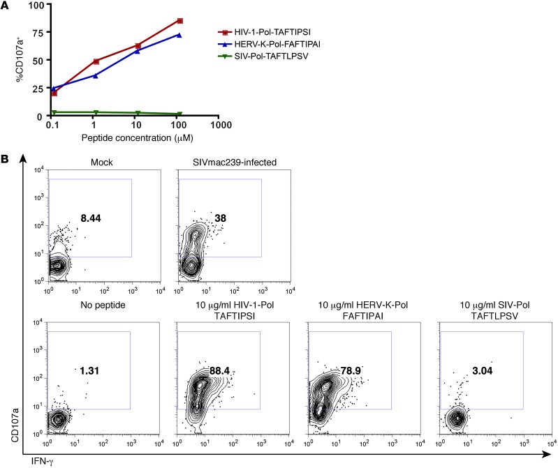
Figure 10. Recognition of SIVmac239-infected cells by a HERV-K(HML-2)-Pol/HIV-1-Pol cross-reactive T cell clone.
A HERV-K(HML-2)-Pol–specific CD8+ T cell clone was isolated from an individual with early (<1 year) HIV-1 infection. (A) This clone was cultured with an autologous BLCL pulsed with the indicated concentrations of the HIV-1-Pol peptide TAFTIPSI, the HERV-K(HML-2)-Pol peptide FAFTIPAI, or the SIVmac239-Pol peptide TAFTLPSV in the presence of PE-conjugated anti-CD107a antibody and brefeldin A. Shown are flow cytometry data depicting the percentage of CD107a+ (degranulating) cells, gated on viable CD8+ clone cells. (B) The HERV-K(HML-2)-Pol–specific CD8+ T cell clone was cultured with HLA-B*51+CD4+ T cells that had been infected with SIVmac239 or maintained as mock-infected controls (top row) or infected with autologous BLCLs pulsed with the indicated peptides (bottom row). Shown are flow cytometry data, gated on CD8+ clone cells and displaying IFN-γ (x axis) by CD107a (y axis). Numbers represent percentages of CD107a+ cells.

