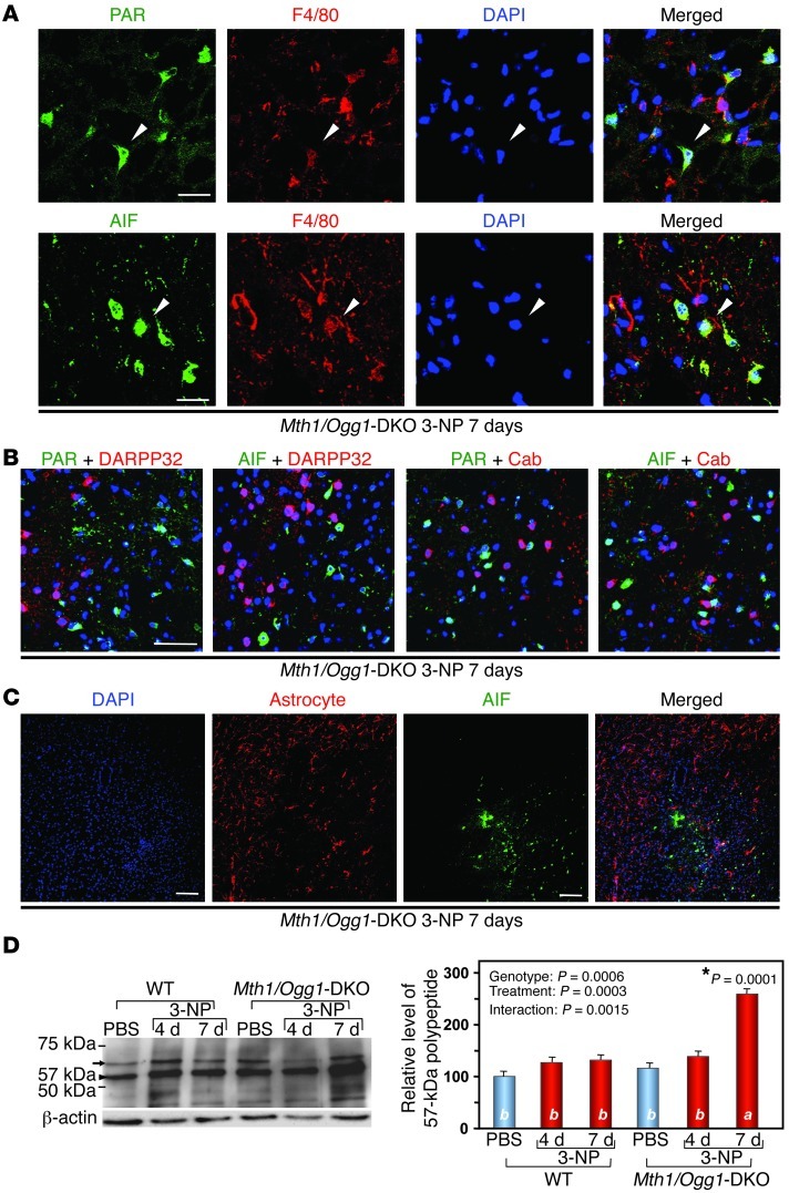
Figure 5. 3-NP induces PAR and AIF accumulation in striatal microglia in Mth1/Ogg1-KDO mice.
(A) Microglia in the DKO striatum exhibited IRs for PAR/AIF. Scale bars: 20 μm. (B) PAR and AIF were not accumulated in MSNs after 3-NP exposure. PAR/AIF, DARPP32 and calbindin (Cab), and DAPI were detected. Scale bar: 50 μm. (C) AIF was not accumulated in astrocytes. Scale bar: 50 μm. (D) 3-NP enhanced processing of AIF in DKO striatum. A 67-kDa mitochondrial form of AIF (arrow) and a 57-kDa nuclear form (arrowhead) are shown (left). Relative levels of the 57-kDa polypeptides are shown in a bar graph (right) (n = 3). In D, data are shown as LS means ± SEM. Levels not connected with the same letter are significantly different (Tukey’s HSD test). *P, compared with the corresponding WT samples.

