Figure 6.

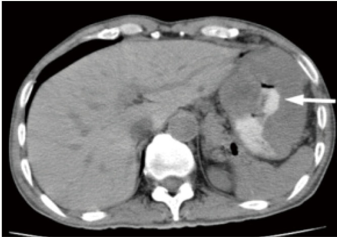
A 52-year-old female with malignant gastric GIST. Axial unenhanced CT image shows an extraluminal mass with necrosis cavity and communication with gastric luminal. Oral contrast materials is seen in the necrosis cavity (arrow)

A 52-year-old female with malignant gastric GIST. Axial unenhanced CT image shows an extraluminal mass with necrosis cavity and communication with gastric luminal. Oral contrast materials is seen in the necrosis cavity (arrow)