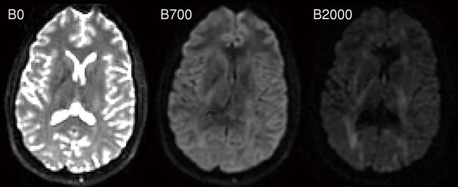
Figure 4.

Diffusion-weighted imaging with different degrees of weighting. The image on the left is a T2-weighted EPI image with no diffusion-weighting (b=0 s·mm-2), the middle image has a modest degree of diffusion-weighting (b=700 s·mm-2) and the right hand image has a high degree of diffusion-weighting (b=2,000 s·mm-2). The diffusion gradients have been applied in the x-direction (left-right). Note the high attenuation in CSF where the diffusion coefficient is high and the lower attenuation in white matter structures such as the internal capsule running perpendicular to the diffusion gradient direction
