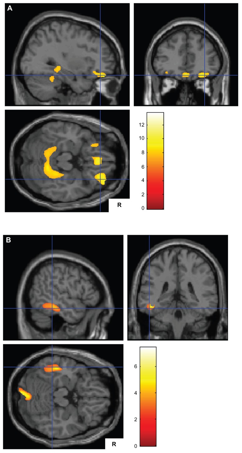
Figure 1.
(A) Smaller gray matter volumes in patients with Alzheimer’s disease and delusions (n = 18) than in those without delusions (n = 35) on both sides of the parahippocampal gyrus, both sides of the orbitofrontal cortex, and both sides of the medial frontal gyrus ventromedial prefrontal cortex. (B) Smaller gray matter volumes in patients with Alzheimer’s disease without delusions (n = 35) than in those with delusions (n = 18) in the left inferior temporal gyrus and the right cerebellum.
Note: The Talairach coordinates are shown in Tables 2 and 3.

