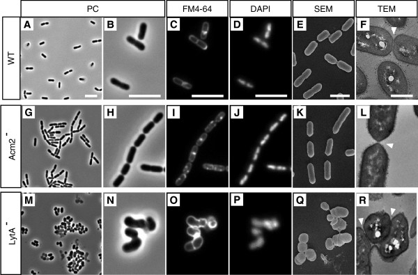
Figure 2.
Cell morphology of L. plantarum and its mutant derivatives. NZ7100 WT (controls; A, B, C, D, E and F), Acm2- (G, H, I, J, K and L) and LytA-(M, N, O, P, Q and R) mutant strains are presented. Micrographs obtained by phase-contrast (PC) optical microscopy (A, B, G, H, M and N), FM4-64 staining (C, I and O), DAPI staining (D, J and P), scanning electron microscopy (SEM) (E, K and Q) and transmission electron microscopy (TEM) (F, L and R) are shown. Cells for microscopy were grown in MRS medium at 28°C and collected in exponential growth phase. The arrows indicate the septum of dividing cells. Bar scales, 5.0 μm for PC, FM4-64 and DAPI; 5.0 nm for SEM and TEM.

