Figure 6.
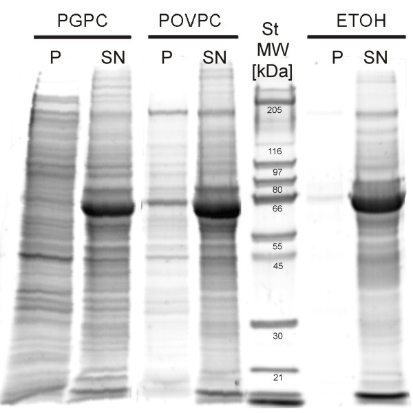
Protein pattern of apoptotic blebs. RAW264.7 cells were incubated with 50 μM PGPC or POVPC or 1% (v/v) EtOH under low serum conditions (0,1% FCS) for 18 h. Membrane vesicles were isolated by ultracentrifugation and resuspended in PBS. Proteins of pellets (P) and supernatants (SN) obtained after ultracentrifugation were separated by SDS-PAGE as described in materials in methods section. Vesicles produced by POVPC and PGPC show slightly different protein patterns. More vesicles protein is released under the influence of PGPC.

