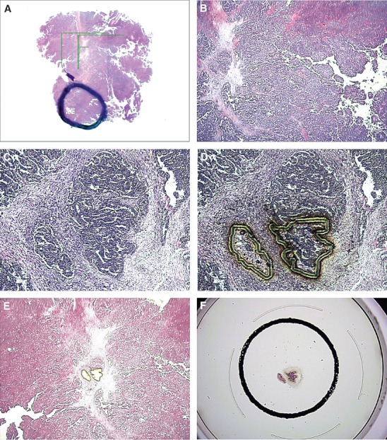
Fig. 1.

Microdissection of HE-stained ovarian cancer tissue slide. (A) overview of tissue slide, with ring marking area used for microdissection. (B) 20× magnified region of interest. (C) 100× region of interest with two cancer cell islets. (D) Laser cutting of region of interest. (E) 20× region of interest after laser capture microdissection. (F) Laser capture microdissected cells on cap, ready for subsequent analysis (real-time PCR).
